Jul 24, 2026
•5 minutes read
•Author: MINDBODYFACE
ANATOMY OF THE FACIAL MUSCLES

Facial muscle anatomy is the study of the muscles that move the skin, shape expression, and support communication. Unlike the larger muscles of the body, facial muscles are thin, closely attached to the skin, and built for precision rather than force.
Understanding anatomy of facial muscles matters because these muscles do more than create a smile or a frown. They help us speak, react, signal emotions, and show age-related changes in the face.
WHAT IS FACIAL MUSCLE ANATOMY AND HOW IT IS STRUCTURED
Facial muscle anatomy refers to the arrangement, origin, insertion, and function of the muscles of the face. The system is usually studied through superficial anatomy, innervation patterns, and how each muscle pulls on skin and soft tissue.
What makes facial anatomy explained differently from limb anatomy is the direct connection between muscle and skin. In many facial muscles, contraction changes expression immediately because the muscle fibers insert into the dermis rather than into bone alone. This is why even a small contraction can alter the visible shape of the face.

Facial musculature is also studied as part of a biomechanical unit: muscle, connective tissue envelope, and skin working together. Age, tension, and tissue quality affect all three layers at once.
FACIAL MUSCLE GROUPS AND OVERALL STRUCTURE
The face contains a large number of small muscles, often divided into facial expression muscles and chewing muscles. The expression muscles are the ones most people think of first, while the mastication group includes the jaw muscles that move the mandible.
The overall structure of facial muscle groups is layered. Some sit superficially under the skin, while others lie deeper around the mouth, eyes, and jaw. This layering creates the visible facial muscle structure that allows fine movement across the brow, cheeks, lips, and chin.
Typical facial muscle groups include the frontalis, orbicularis oculi, zygomaticus major and minor, orbicularis oris, buccinator, mentalis, platysma, masseter, and temporalis. Each one has a narrow task, but together they create coordinated facial motion.
MUSCLES RESPONSIBLE FOR FACIAL EXPRESSIONS
The muscles responsible for facial expressions are called mimetic muscles, because they mimic emotional and social states. These are the muscles that create smiling, frowning, squinting, lip pressing, and eyebrow movement.
The mimetic muscles of the face are innervated by the facial nerve, cranial nerve VII. This is one of the main reasons facial expression muscles are treated as a separate system from chewing muscles, which are controlled by the trigeminal nerve. That difference in innervation helps explain why facial movement is so expressive and precise.

A smile depends mainly on the zygomaticus muscles, the orbicularis oculi tightens the eyes during squinting and genuine smiling, and the frontalis lifts the brows in surprise or alertness. A frown relies more on the corrugator, procerus, and depressor muscles around the brows and mouth. The anatomy of facial expressions is therefore not abstract — it is a map of repeated muscle actions.
FACIAL MUSCLES DIAGRAM AND VISUAL UNDERSTANDING
A facial muscles diagram is one of the clearest ways to learn the system because the muscles overlap and can be hard to see in isolation. Imaging and anatomical illustrations help separate the groups by origin, insertion, and line of pull.
The classic diagram of the face typically shows the frontalis, orbicularis oculi, zygomaticus major, risorius, orbicularis oris, mentalis, buccinator, and platysma. In some diagrams, deeper or functionally important muscles are shown as well, especially when the goal is to understand expression mechanics rather than memorise names.
Visual mapping matters because facial muscles are small and interwoven. A good diagram turns the facial anatomy into a readable system instead of a list of isolated terms.
FACIAL MUSCLE FUNCTION AND MOVEMENT MECHANICS
Facial muscle function is based on small contractions that pull on skin and soft tissue. Most facial muscles shorten in one direction only, so movement comes from combinations rather than from one muscle acting alone.
For example, a smile is not created by one muscle. It is the result of several muscles coordinating lift, pull, and soft-eye engagement. A frown works the same way in reverse: brow adduction, eye narrowing, and lower-face tension create the expression.
The mechanism is simple but elegant. Muscles contract, the connective tissue transmits force, skin moves, and the expression appears instantly. Because the face is a social signal system, even small changes in muscle tone can change how a person is perceived.
FACIAL MUSCLES AND AGING
Facial muscles and aging are linked through tone, coordination, and the condition of surrounding tissues. Age-related changes occur not only in the skin, but also in the muscle and connective tissue envelope that supports it.
In the upper face, studies show that older participants had reduced skin elasticity and lower frontalis activity, which changed how the forehead and brow moved. In the cheek area, facial sagging was associated with reduced skin elasticity, increased fat mass, and weaker mimetic muscle function. That means aging is not just a skin issue — it is also a muscle and support-tissue issue.

As facial tone changes over time, expression can look heavier, flatter, or more tired. The face may also rely on compensatory patterns, where some muscles overwork while others become less active. This is why facial aging patterns often show up first in movement, then in rest.
CAN SLEEPING ON ONE SIDE CHANGE YOUR FACE? SLEEP LINES, ASYMMETRY, AND FACIAL COMPRESSION
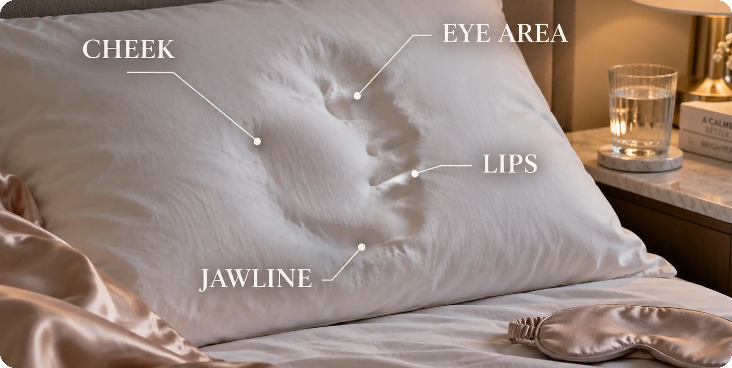
Can sleeping on one side change your face? It can temporarily change how your face looks in the morning. One cheek may seem puffier, one eye more swollen, and one side may show deeper pillow marks. Over many years, repeated pressure may contribute to sleep lines in aging skin, but side sleeping alone does not […]
Author:MINDBODYFACE
 8
8 0
0
UNEVEN SMILE: WHY ONE SIDE OF THE MOUTH SITS HIGHER AND WHAT FACIAL TENSION HAS TO DO WITH IT
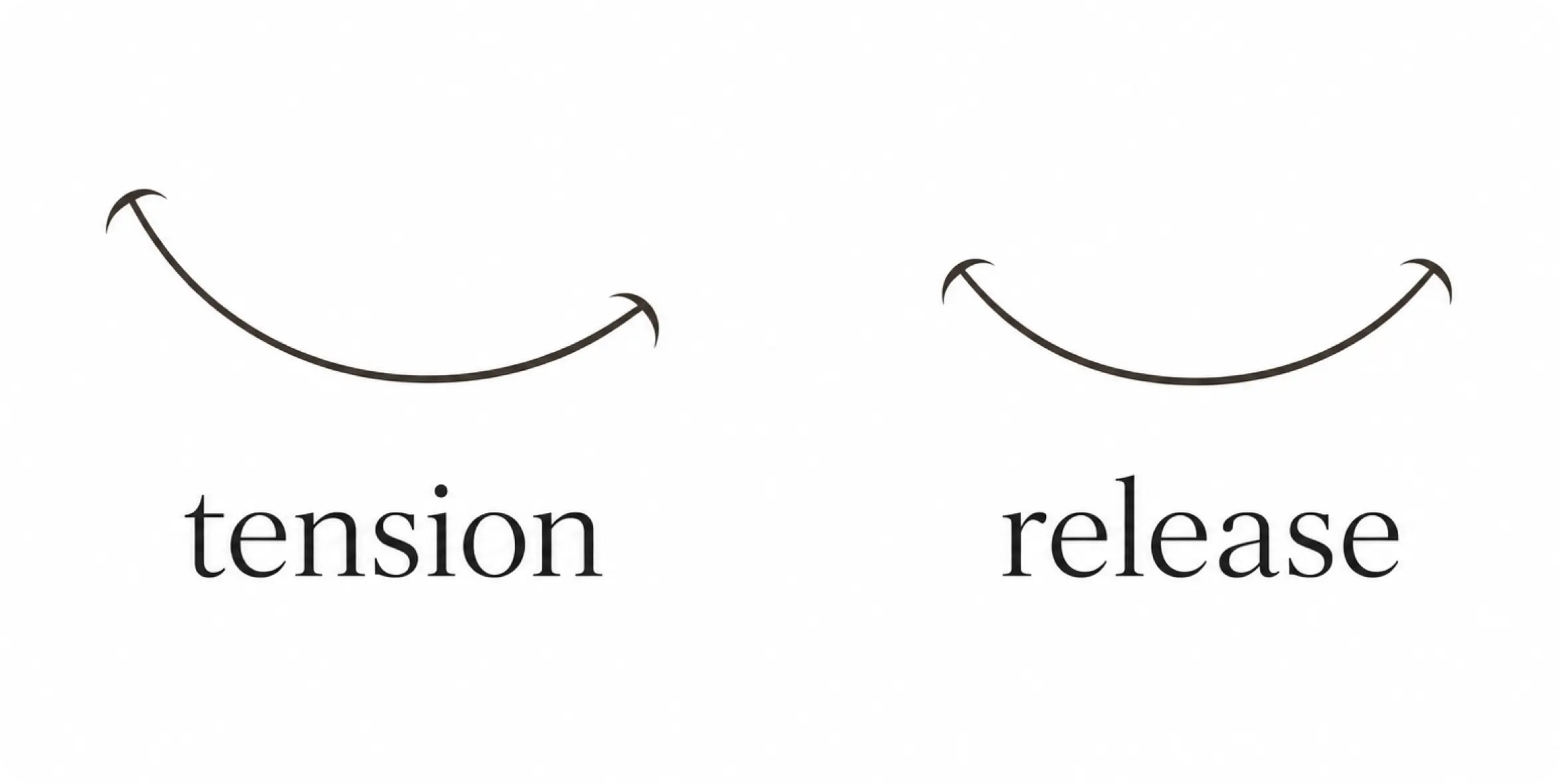
An uneven smile can be one of those details you only notice in photos or videos. One corner of the mouth lifts more. One side of the upper lip looks higher. One cheek seems to move first, while the other appears quieter or tighter. A little facial asymmetry is normal. Almost no one has a […]
Author:MINDBODYFACE
160
DIMPLED CHIN AND MENTALIS TENSION: WHY THE CHIN LOOKS UNEVEN AND HOW TO RELAX IT
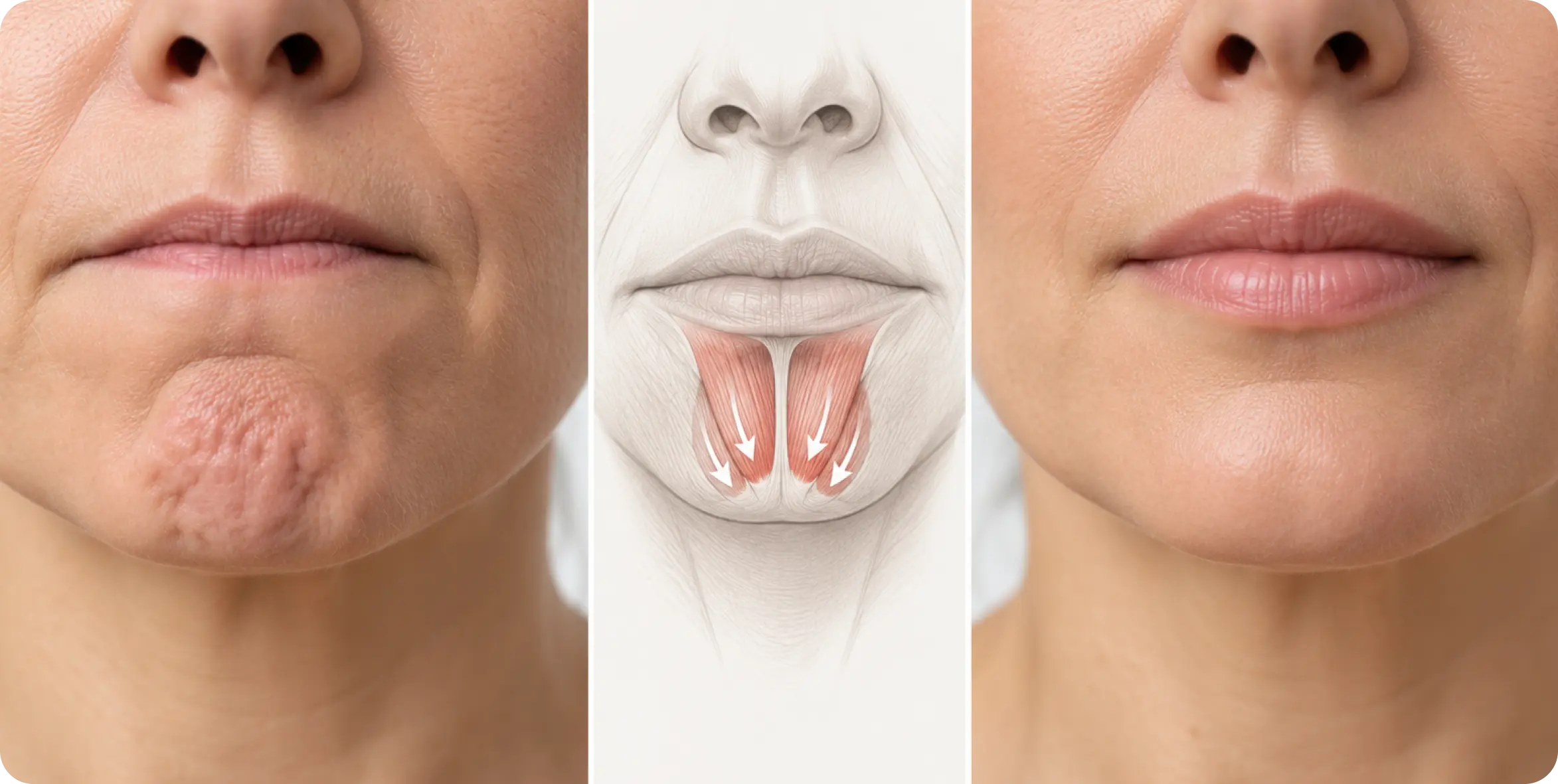
A dimpled chin can make the lower face look tense even when you are not upset. The skin may appear bumpy, gathered, or uneven when you talk, smile, concentrate, swallow, or press your lips together. For some people, this texture appears only during expression. For others, it remains visible at rest. The common reason is […]
Author:MINDBODYFACE
140
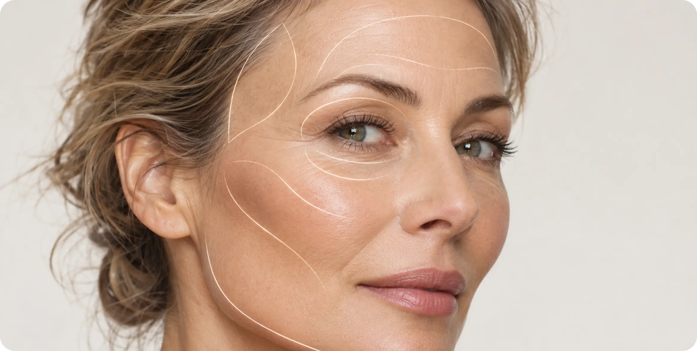
HOLLOW TEMPLES: WHY TEMPLES LOSE VOLUME AND HOW FACIAL AGING CHANGES THIS AREA
Hollow temples are the indentations that can appear at the sides of the forehead, between the outer eyebrow, hairline, and upper cheekbone. Temple hollowing can make the upper face look narrower, more angular, or more tired—even when your skin is smooth and your cheeks still have volume. This area is easy to overlook. People often […]
Author:MINDBODYFACE
260


MINDBODYFACE LLC
1021 E Lincolnway Suite 8342, Cheyenne, WY 82001
+16282892382
All services and information on this website are for educational purposes only and do not constitute medical advice or treatment -